
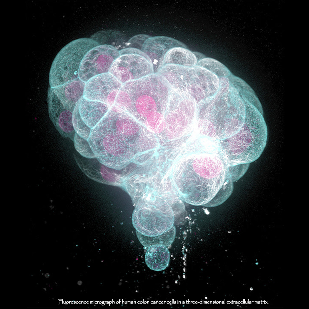
 Fluorescence micrograph of human colon cancer cells in a three-dimensional extracellular matrix. This environment mimics physiological tissue, and the cells organize into cancer organoids. Such 3D organoid cultures are used by researchers to understand the molecules and mechanisms involved in tissue formation, and in the case of cancer to understand pathological cell behavior and to develop new therapies. These cells were stained by immunofluorescence for two components of the cell cytoskeleton: Filamentous actin (cyan) that highlights microscopic cell surface structures and cell-cell contacts, and microtubules (white) that form a transport system inside cells. In addition, the DNA in cell nuclei appears red.
Tags: #cells #biology #cancer #health #cell #3D #bio #nerds #geek #micro #microscopic #macro #photooftheday #imageoftheday #imageoftheweek #sciencebitch #technology #tech #medicine #picoftheday #picoftheweek #follow4follow #f4f #followforfollow #l4l
CREDIT: DR TORSTEN WITTMANN – SPL
YOU MAY LIKE: Personalized Cancer Treatment 2.0: New Combination Strategy Developed


